Your shopping cart is empty!


DOCTOR INFORMATION
Abdominal Examination (OSCE)
Abdominal Examination
Introduction
- Greet the patient and introduce yourself

- Confirm patient details
- Briefly explain the examination in a patient friendly manner
- Get patient consent
- Set the head of the bed to a 45° angle 📐
- Wash hands ✋
- Expose patient’s abdomen and lower legs
- Check the patient is not in any pain
General inspection of the patient
Identify any clinically relevant signs:
 Age: e.g. IBD more common diagnosis in younger patient whereas malignancy and chronic liver disease is more common in older patients
 Pain: examine the area where the pain is
Confusion: end stage liver disease symptom
Pallor: anaemia
Jaundice: high bilirubin levels associated with liver pathology
Scars: prior surgery
Hyperpigmentation: darkening of the skin due to haemochromatosis
Hernias: coughing can make them more visible
Oedema: swollen limbs or abdomen due to liver cirrhosis
Cachexia: muscle loss due to malignancy or advanced liver failure
Identify any objects or equipment that may be relevant:
Prescriptions: recent medications 💊
Mobility aids ♿
Feeding tubes
Fluid balance: indicate fluid overload or dehydration
Vital signs: reflect past and present clinical status
Surgical drains: record the type and location
Stoma bag: record the location
Other medical equipment
Hands
Inspection of hands
Inspect the palms ✋:
Pallor: anaemia
Dupuytren’s contracture
Palmar erythema: red palm of hand, can indicate liver disease
Inspect the nails ✋:
Leukonychia: hypalbuminaemia can cause the nail bed to whiten
Koilonychia: anaemia can present with ‘spoon shaped’ nails
Assess for finger clubbing ✋:
- Ask the patient to put the nails of their index fingers back to back
- If you cannot see a diamond shaped window, the patient has ‘finger clubbing’
The normal angle between the nail and the nail bed is lost due to soft tissue in the finger-tip swelling
Can indicate: coeliac disease, IBD, lymphoma of the gastrointestinal tract and liver cirrhosis
Assess for Asterixis ✋:
- Ask the patient to put both arms out in front of them, bending their hands backwards at the wrist joint and holding for 30 seconds
- Observe for hand ‘flapping’
Asterixis, or ‘flapping tremor’ is identified by the hands flapping
Can indicate: hepatic encephalopathy, uraemia, or CO2 retention
Palpation of the hands
Assess the temperature:
Symmetrical warmth indicates the patient is healthy
Cold hands indicate poor peripheral perfusion ❄
Assess the radial pulse:
- Use your middle and index fingertips to palpate
- Assess the rate and rhythm
Assess for Dupuytren’s contracture:
- Palpate the palm
- Identify thickened, ‘cord-like’ bands of palmar fascia
The thickening of the palmar fascia, producing ‘cords’ will cause finger and thumb deformities
Arms and axillae
Inspect the arms:
Needle track marks: indicative of intravenous drug use which can increase hepatitis risk
Bruising: indicative of clotting abnormalities caused by liver disease
Excoriations: scratch marks which may indicate cholestasis
Inspect the axillae:
Hair loss: due to iron deficiency anaemia or malnutrition
Acanthosis nigricans: thickened or darkened skin due to insulin resistance or gastrointestinal malignancy
Face
Inspect the eyes:
Jaundice: visible in the upper region of the sclera
Perilimbal injection: inflammation of the conjunctiva region adjacent to the iris, can be indicative of IBD
Corneal arcus: white/grey/blue ring in the peripheral cornea, in patients <50 indicates hypercholesterolaemia
Kayser-Fleischer rings
Conjunctival pallor: indicates anaemia
Xanthelasma: raised, yellow, cholesterol rich deposits on the area around the eye which indicate hypercholesterolaemia
Inspect the mouth:
Oral candidiasis: fungal infection, identified by white mouth, caused by immunosuppression
Hyperpigmented macules: polyps in the gastrointestinal tract caused by the genetic, Peutz-Jeghers syndrome
Glossitis: enlarged tongue 👅, caused a deficiency in B12, iron and folate
Aphthous ulceration: round or ovular ulcers on the mucosal membranes, which can also indicate B12, iron and folate deficiency
Angular stomatitis: inflammation of the corners of the mouth caused by a variety of things such as iron deficiency
Neck
Metastatic intrabdominal malignancy can be identified by a swollen Virchow’s node (left supraclavicular lymph node)
Oesophageal cancer can be identified when the right supraclavicular lymph node is swollen
Palpate for lymphadenopathy:
Palpate on both sides to identify swelling
Chest
Hair loss: can be caused by increased oestrogen or malnourishment
Spider naevi: lesions with a red centre and fine red lines extended from it ‘like a spider’, due to increased oestrogen levels, >5 indicate liver cirrhosis
Gynaecomastia: male breast tissue enlarges due to increased oestrogen or medication such as digoxin/ spironolactone
Abdomen
Hernias: assess for protrusions when coughing
Scars: indicate previous surgery
Striae: ‘stretch marks’ caused by rapid growth or skin stretching
Abdominal distension: caused by the 6F’s (foetus/faeces/fat/fluid/flatus)
Cullen’s sign: bruising around the umbilicus indicates late stage pancreatitis
Caput medusae: swollen paraumbilical veins and hypertension of the portal vein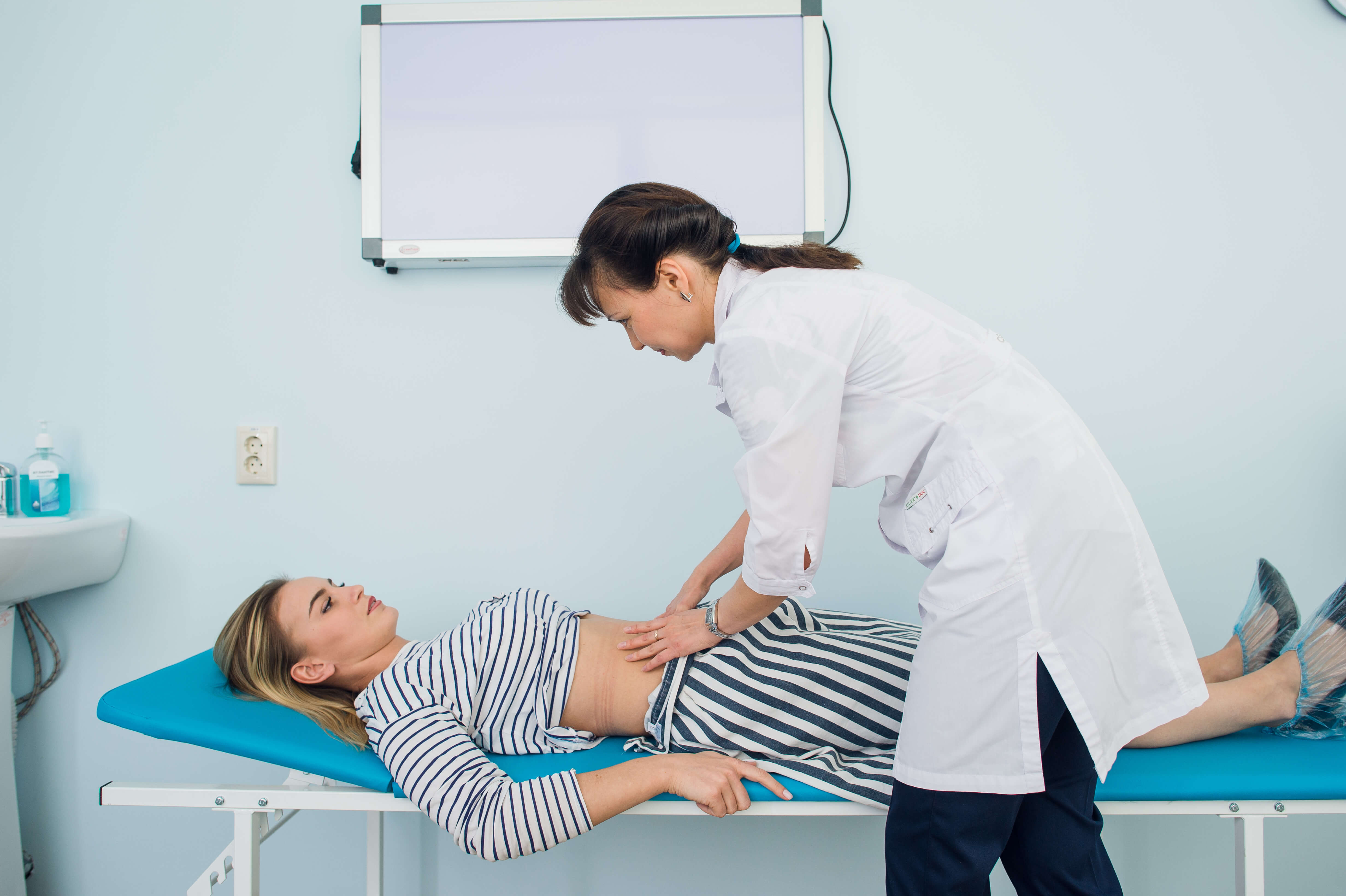
Grey-Turner’s sign: bruised flanks indicating haemorrhagic pancreatitis
Assess any stomas present:
Contents: stool/urine
Spout: no spout in ileostomies/urostomies, spout present in colostomies
Location: indicates type of stoma (right iliac fossa = ileostomies and urostomies, left iliac fossa = colostomies)
Palpation of the abdomen
Prepare for palpation:
- Lay patient flat on bed
- Check for abdominal pain
- Identify pain by watching their face when palpating
Light palpation:
Assess for signs of pathology as you lightly palpate the 9 abdominal regions:
Masses
Tenderness
Guarding: involuntary tensing
Rovsing’s sign: pain in right iliac fossa when palpating the left, can indicate peritonitis
Rebound tenderness: slow compression and quick release of the abdominal wall causes pain which can indicate peritonitis
Deep palpation:
Apply more pressure and assess for signs of pathology as you palpate the 9 abdominal regions:
Mobility: distinguish between superficial masses and those attached to other structures
Location: record which region the mass is present
Pulsatility: pulsatile masses, associated with vascular origins
Consistency: smooth/soft/hard etc.
Size/shape: approximately measure the size of the mass
Liver palpation:
- Palpate the right iliac fossa as the patient inhales deeply to feel for the liver edge
- Repeat, moving 1 to 2 cm upwards each time until you reach the right costal margin
- If you identify the liver edge, record the following:
- Position: greater than 2cm beneath the costal margin indicates hepatomegaly
- Tenderness: can indicate hepatitis or cholecystitis
- Consistency: if the liver edge feels nodular, it can indicate cirrhosis
- Pulsatility: can indicate tricuspid regurgitation
Hepatomegaly causes:
Hepatitis
Leukaemia
Haemolytic anaemia
Wilson’s disease
Glandular fever
Hepatocellular carcinoma
Primary biliary cirrhosis
Hepatic metastases
Haemochromatosis
Myeloma
Tricuspid regurgitation
Gall bladder palpation:
If you can palpate the gall bladder, it suggests it is enlarged due to biliary flow obstruction/infection
- Palpate at the right costal margin, in the mid-clavicular line
- If you identify a motile round mass, it suggests the gall bladder is enlarged
- Pain suggest cholecystitis (Murphy’s sign)
- Murphy’s sign is identified when you palpate the fall bladder as the patient inhales deeply and they suddenly stop mid-breath because they are in pain
- No pain suggests pancreatic cancer
Spleen palpation:
- Palpate the right iliac fossa as the patient inhales deeply to feel for the splenic edge
- Repeat, moving 1 to 2 cm towards the left costal margin
- If you identify the splenic edge, it is suggestive of splenomegaly
Splenomegaly causes:
Glandular fever
Haemolytic anaemia
Portal hypertension caused by liver cirrhosis
Splenic metastases
Congestive heart failure
Kidney Balloting:
- Place your left hand ✋ under the back, standing on the opposite side
- Place your right hand ✋ just below the right costal margin
- Push your right hand ✋ down and your left up as the patient inhales deeply
- If you ballot the kidney, record its consistency and size
- Repeat for the left kidney
Enlarged kidney causes:
Unilaterally enlarged: renal tumour
Bilaterally enlarged: polycystic kidney disease or amyloidosis
Aorta palpation:
- Palpate just above the umbilicus in the midline using both of your hands
- Record how your fingers move:
- Outwards: indicates expansile mass
- Superiorly: normal
Further tests are require before diagnosis
Bladder palpation:
- Ask the patient to use the toilet before beginning
- Palpate in the suprapubic area to identify if it is palpable and hence distended
Abdominal percussion
Liver percussion:
- Percuss the right iliac fossa
- Repeat, moving 1 to 2 cm towards the right costal margin until the percussion note becomes dull rather than resonant (indicates border of lower liver)
- Continue to percuss upwards, 1 to 2 cm at a time until the percussion notes changes from dull to resonant (indicates upper liver border)
- Approximate the size of the liver
Spleen percussion:
- Percuss the right iliac fossa
- Repeat, moving 1 to 2 cm towards the left costal margin, until the percussion note becomes resonant rather than dull (indicates location of spleen)
- If you identify the spleen it is suggestive of splenomegaly
Bladder percussion:
- Percuss in the midline of the umbilical region, moving downwards towards the pubic symphysis
- When the percussion note becomes dull, you have identified the upper border of the bladder
Shifting dullness:
- Percuss from the umbilical region, upwards towards the left flank
- A dull percussion note can indicate ascitic fluid in the flank
- Ask the patient to role onto their side for 30 seconds ⏱ (keep your fingers in the position where the note became dull)
- Repeat percussion, the percussion note should now be resonant to confirm ascites presence
Abdominal auscultation
Bowel sounds 👂:
Auscultate in ≥2 abdominal areas
Normal bowel sounds: gurgling
Absent bowel sounds: suggestive of ileus (malfunctioning peristalsis disrupts the intestine functioning)
Tinkling bowel sounds: bowel obstruction
Bruit identification:
Identify vascular bruits (indicate turbulent blood flow) by auscultating over the aorta and renal arteries
Aortic bruits: 1 to 2 cm above the umbilicus, can indicate abdominal aortic aneurysm
Renal bruits: 1 to 2 cm above the umbilics, but slightly to the side of the midline on both sides, can indicate renal artery stenosis
Legs
Identify pitting oedema on the lower legs, can indicate hypalbuminaemia
Completion
- Tell the patient the examination is complete, and thank them
- Wash hands ✋
- Summarise what the examination revealed
Summary:
- Greet the patient and briefly explain the examination
- Inspect the patient to identify any clinically relevant signs
- Inspect the patient's arms, axillae, face and mouth
- Palpate the neck for lymphadenopathy
- Inspect the chest and abdomen for any clinical features
- Assess any stomas present
- Palpate, percuss and auscultate the abdomen
- Assess for pitting oedema on the lower legs
- Complete the examination by thanking the patient

Related Articles
This step by step guide is designed to take you through the hernia examination in OSCEs.