DOCTOR INFORMATION
GALS (Gait, Arms, Legs, Spine) Examination (OSCE)
Introduction
 Greet the patient and introduce yourself
 Confirm patient details
 Briefly explain the procedure in a patient friendly manner
 Get patient consent ✅
 Expose the patient as required
 Wash hands ✋
Screening Questions
- Do you suffer from pain/stiffness in muscles/joints/back❓
- Do you struggle to dress yourself independently❓
- Do you have any difficulty using stairs❓
Gait
Observe the patient as they walk to the end of the room and back:
 Gait cycle: identify abnormalities 🚶
 Waddling gait: indicates weakness of the hip abductor muscles on both sides, associated with myopathies
 Trendelenburg’s gait: indicates weakness of the hip abductor muscles on one side, due to L5 radiculopathy or superior gluteal nerve lesion
 Leg length: differences can indicate joint pathology 📏
 Limp: can indicate joint pain/weakness
 Slow turning: can indicate joint restrictions
 Footwear: unequal wearing of the sole can indicate an abnormal gait 🚶
 Movement: reduced range of movement indicates chronic joint pathology
Normal gait cycle:
- Heel makes contact with floor 🚶
- Foot becomes flat and weight is transferred onto it
- Weight balanced on flat foot’s leg
- Heel lifted off floor
- Toes lifted off floor
- Foot swings forward and cycle begins again

Inspection
Identify any clinically relevant signs:
 Scars: indicate prior surgery
 Psoriasis: scaly plaques on extensor surfaces, can indicate psoriatic arthritis
 Obesity: significant joint pathology risk factor ❌
 Muscle wastage: indicates lower motor neuron injury or disuse atrophy
Identify any clinically relevant objects or equipment:
 Prescriptions: indicate recent medications 💊
 Aids/adaptations: wheelchair/walking aids/slings/splints ♿
Inspect the patient from an anterior aspect in a standing position:
 Scars: indicate prior surgery or trauma
 Swollen joints: identify unilateral swelling which can indicate effusion, septic arthritis or inflammatory arthropathy
 Erythema of joints: indicates active inflammation which can indicate septic arthritis or inflammatory arthropathy
 Posture: asymmetry can indicate scoliosis or pathology of joints
 Deformity of the valgus joint: knees knocking together
 Deformity of the varus joint: bowlegged appearance
 Muscle bulk: upper and lower asymmetry can indicate lower motor neuron injury or disuse atrophy
 Big toe: identify medial or lateral angulation 👣
 Elbow extension: identify cubitus valgus (carrying angle above within 5-15°), caused by congenital deformity or elbow joint trauma or cubitus varus “gunstock deformity” (carrying angle below 5-15°), caused by supracondylar fracture of the humerus
 Lateral pelvic tilt: caused by difference in leg lengths, scoliosis or weak hip abductor muscles
Inspect the patient from a lateral aspect in a standing position :
 Arched foot: inspect for flat feet or a raised foot arch 👣
 Lumbar lordosis: loss of normal inward curve of the spine’s lumbar region indicates sacroiliac joint disease
 Cervical lordosis: hyperlordosis suggests chronic degenerative joint disease
 Hyperextension of knee joint: caused by ligamentous damage or hypermobility syndrome
 Thoracic kyphosis: normal range is 20-45°, higher indicates Scheuermann’s disease 📐
Inspect the patient from a posterior aspect in a standing position:
 Deformity of valgus joint: foot turned outwards 👣
 Deformity of the varus joint: foot turned inwards 👣
 Spinal alignment: lateral curvature of spine indicative of scoliosis
 Thickened Achilles tendon: Achilles tendonitis
 Muscle bulk: asymmetry between upper and lower limbs can be caused by lower motor neuron injury or disuse atrophy
 Iliac crest alignment: misalignment can indicate weakness of hip abductor muscles or or difference in leg length
 Popliteal swellings: indicates possible Baker’s cyst or popliteal aneurysm
Arms
Assess compound Movements:
- Ask patient to put hands behind head and pint elbows to sides ✋
- Restricted movement range indicates pathology of shoulder/elbow
- Excessive movement range can suggest hypermobility
- Ask patient to hold their arms out in front of their body with their palms facing the floor and their finger stretched out
- Assess dorsum for asymmetry/deformity/joint swelling
- Assess nails for signs indicative of psoriasis
- Ask patient to turn hands over ✋
- Restricted movement indicates pathology of wrist/elbow
- Ask patient to form fist with hand ✋
- Inability to do so can be due to swollen joints/deformities
- Ask patient to squeeze your fingers, assessing strength of grip
- Low grip strength can be caused by pain/ lower motor neuron lesions
- Ask patient to touch each finger with thumb of same hand ✋
- Inability may be due to inflammation or the small joint may have joint contractures
Squeeze the Metacarpophalangeal joint:
 Assess patient for discomfort when you squeeze the joints gently
 Tenderness indicates active inflammatory arthropathy
Legs
Assess passive movement, controlled by you:
 Passive knee flexion: support knee and flex leg as far as possible before patient experiences discomfort (normal 0-140°) 📐
 Passive knee extension: a patient laying flat on the bed is showing normal knee extension, assess for hyperextension by holding above their ankle, then lifting the leg upwards identifying hyperextension >10° 📐
Passive internal hip rotation:
 Flex hip and knee joint to 90°, then rotate foot laterally (normal 40°) 📐
Squeeze the metatarsophalangeal joint:
 Assess patient for discomfort when you squeeze the joints gently
 Tenderness indicates active inflammatory arthropathy
Tap the patellar:
 Can identify moderate/large knee joint effusion caused by ligament rupture, inflammatory/septic/osteo-arthritis
- Extend the knee fully and slide your hand down the thigh until it reaches the patella’s upper border ✋
- Holding your left hand in that position, assert pressure on the patella with your right hand ✋
- A distinct tap indicates the presence of fluid as the patella bumps against the femur
Spine
 Ask the patient to stand upright
Cervical lateral flexion:
 Ask patient to try to touch their ears to shoulders
 Assess cervical spine lateral flexion
Lumbar flexion:
 Palpate for a range of movements to assess lumbar flexion
- Place 2 fingers between 5 and 10cm apart of the lumbar vertebrae 📏
- Ask patient to touch their toes by bending towards downwards
- Assess whether your fingers move apart
- Ask the patient to stand straight
- Assess whether your fingers move back together
 Hypermobility = ability to place hands flat on the floor ✋
Temporomandibular joint
 Observe as patient inserts 3 of their fingers into their mouth
 Identify deviation or restriction of jaw movement
Completion
 Tell the patient that the examination is now complete ✅
 Thank patient
 Wash hands ✋
 Summarise what the examination revealed
Summary:
- Greet the patient and briefly explain the examination, then perform screening questions
- Observe the patient walking from one side of the room to the other to assess if they have a normal gait cycle
- Inspect the gait, arms, legs and spine to identify anything clinically relevant
- With the patient in a standing position, inspect them from an anterior, lateral and posterior aspect
- Assess the compound movement of the arms and squeeze the Metacarcophalangeal joint
- Assess passive movement of the legs and passive internal hip rotation, squeeze the Metatarsopholangeal joint, and Tap the patellar
- With the patient standing upright, assess cervical lateral flexion, and lumbar flexion
- Assess the Temporomandibular joint
- Complete the procedure by thanking the patient
Related Articles
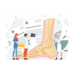
This step by step guide is designed to take you through the ankle and foot examination in OSCEs.
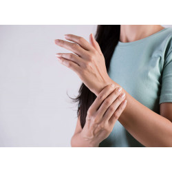
This step by step guide is designed to take you through the hand and wrist examination in OSCEs.
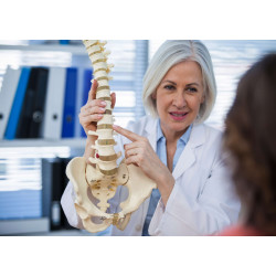
This step by step guide is designed to take you through the Spine Examination in OSCEs.


