Your shopping cart is empty!


DOCTOR INFORMATION
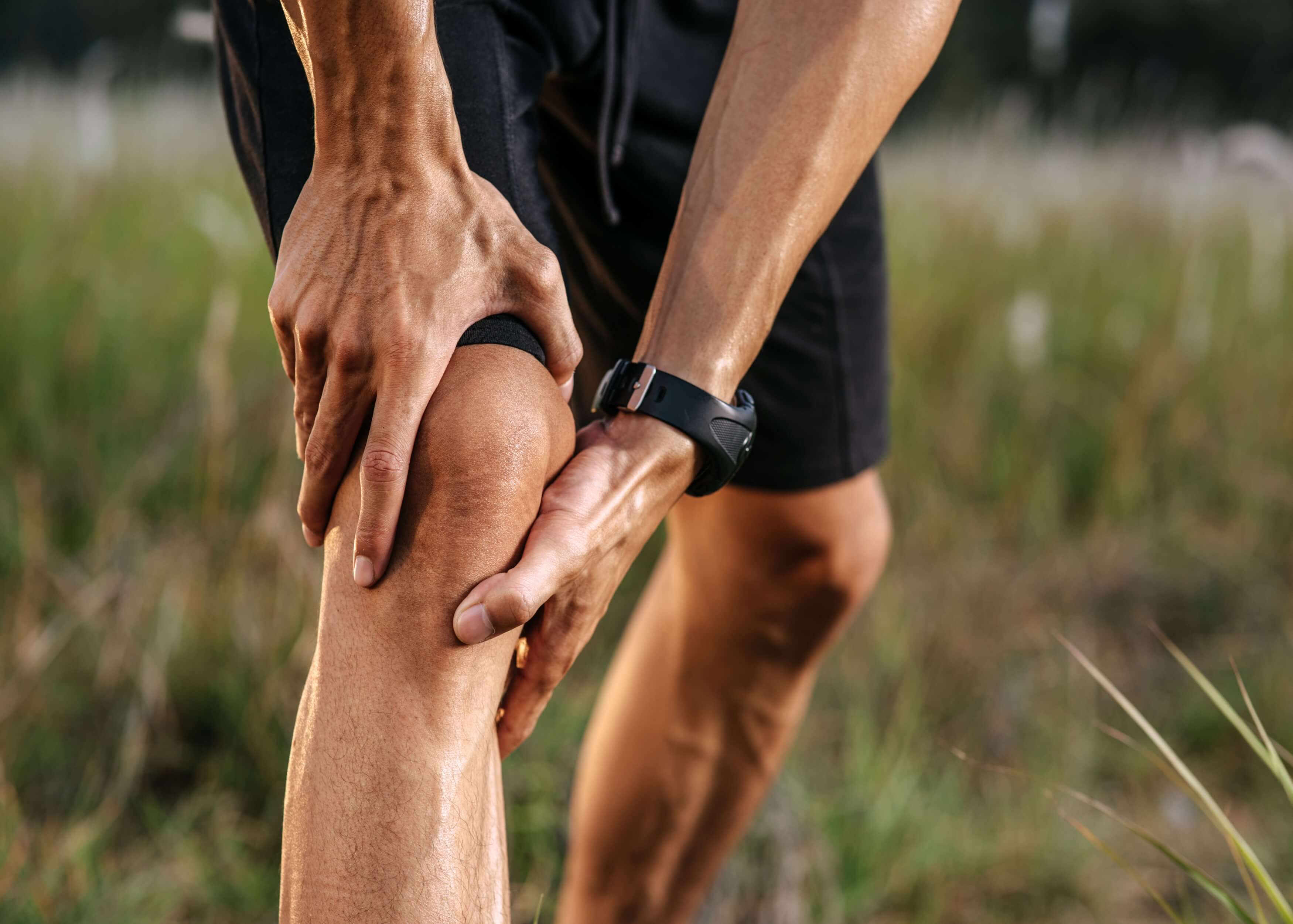
Knee Examination (OSCE)
Introduction
 Greet the patient and introduce yourself
 Confirm patient details
Briefly explain the examination in a patient friendly manner
Get consent ✅
Expose the patient’s legs
Wash hands ✋
Check the patient is not in any pain
Look
Identify any clinically relevant signs:
Scar: indicative of prior surgery 🏨
Obesity: can cause joint pathology
Muscle wastage: indicates disuse atrophy or lower motor neuron injury
Identify any objects/equipment that indicate clinical status :
Prescriptions: indicate recent medications 💊
Walking aids: indicate knee/ hip/ ankle pathology ♿
Inspect the anterior aspect of the knees :
Bruising: indicates trauma/spontaneous haemarthrosis
Scars: indicates trauma/prior surgery 🏨
Swelling: asymmetry indicative of unilateral swelling
Wastage of quadriceps: asymmetry may be due to lower motor neuron lesion or disuse atrophy
Patellar position: non-central position indicates dislocation/subluxation
Varus deformity: tibia turned inwards compared to femur
Valgus deformity: tibia turned outwards compared to femur
Psoriasis plaques: increases psoriatic arthritis risk
Inspect the lateral aspect of the knees:
Fixed flexion deformity: can be caused by contractures due to inflammatory conditions/prior trauma/neurological disease
Extension abnormality: hyperextension of the knee can be caused by cruciate ligament injury
Inspect the posterior aspect of the knees:
Muscle wastage: asymmetry due to lower motor neuron lesion or disuse atrophy
Scars: indicative of prior surgery or trauma 🏨
Popliteal swelling: caused by popliteal aneurysm or Baker’s cyst
Gait
Assess the patient as they walk from one end of the room to the other and back:
Gait cycle: identify abnormalities 🚶
Leg length: identify any differences between the 2 legs, indicative of joint pathology
Limping: indicative of joint pain
High step: can indicate peroneal nerve palsy
Movement range: chronic joint pathology can reduce range of movement
Slow turning: due to joint pathology
Normal gait cycle:
- Heel makes contact with floor 🚶
- Foot becomes flat and weight is transferred onto it
- Weight balanced on flat foot’s leg
- Heel lifted off floor
- Toes lifted off floor
- Foot swings forward and cycle begins again 🔄
Assess the knees when the patient is in the supine position:
Bruising
Scars
Swelling
Asymmetry of knee joint
Wastage of quadricep muscle
Patellar position abnormality
Fixed flexion deformity
Feel
Assess the temperature of the knee joints for comparison:
High temperature can indicate pathology, especially when in conjunction with tenderness and swelling 🌡
e.g. inflammatory arthritis, septic arthritis, gout/pseudogout
Measure the circumference of the quadriceps:
Wrap a measuring tape, about 20cm above the tibial tuberosity
A significant difference between the 2 legs indicates muscle wastage
Palpate the extended knee:
- Palpation of the patella: stabilise one side of the patella and palpate the other using your fingertip 👉:
Tenderness can indicate injury/patellofemoral arthritis
Apprehension may indicate recurrent patellar dislocation
- Palpation of the patellar ligament:
Tenderness indicates rupture/tendonitis
- Palpation of the medial and lateral joint lines:
Tenderness indicates fracture/collateral ligament injury/meniscal injury
- Palpation of the quadriceps tendon:
Tenderness indicates tendonitis/rupture
Joint effusion assessment:
May be caused by ligament rupture, inflammatory arthritis, septic arthritis and osteoarthritis
Patellar Tap test 👉:
- Extend the patient’s knee fully and slide your left hand down the patient’s thigh to the patella’s upper border, emptying the suprapatellar pouch
- Hold your left hand still and press down on the patella with your right fingertips 👉
- If you feel a tap (the patella bumping against the femur), it indicates the presence of fluid
Sweep test: used for joint effusions which are too small to be identified by the tap test
- Lie the patient down and extend their knee fully, repeat step 1 of the tap test 👉
- Stroke the knee joint of the medial side, this will move any fluid over to the lateral side
- Stroke the lateral side of the knee to move the fluid back to the medial side, causing a bulge which indicates the presence of effusion
Palpate the flexed knee:
Flex knee to 90° 📐
- Palpate patella as when extended
- Palpate medial and lateral joint lines as when extended
- Palpate tibial tuberosity to identify bony elevation/tenderness (Osgood-Schlatter disease)
- Palpate head of the fibula to identify fracture indicated by tenderness
- Palpate the popliteal fossa to identify swelling, indicative of a popliteal cyst, or a pulsatile mass which can indicate a popliteal aneurysm
Move
Assess active movement (performed independently):
Active knee flexion: flex knee as far as possible (normal = 0-140° 📐)
Active knee extension: extend knee so leg is flat on bed (normal = 180° 📐)
Assess passive movement (clinician controlled):
Passive knee flexion: flex knee as far as possible (normal = 0-140° 📐)
Passive knee extension: if they are laying flat on bed, patient is exhibiting normal extension
- Hold just above the ankle and raise the leg
- Observe the knee to identify hyperextension (normal = <10° 📐)
Special tests
Assess the cruciate ligaments:
Posterior sag sign: indicates rupture of the posterior cruciate ligament which normally prevent the tibia moving backwards and the femur moving forwards
- Ask patient to flex knee to 90°, placing foot flat on bed 📐
- Inspect the lateral aspect
Anterior drawer test: to assess anterior cruciate ligament integrity
- Lay patient on bed with knee flexed to 90° and relaxed 📐
- Wrap your fingers behind knee joint, stabilising your hand with your forearm on their lower leg
- Place your thumbs over the tibial tuberosity
- Pull tibia anteriorly, feel for significant movement of the tibia on the femur which indicates rupture or laxity of the ligament
Posterior drawer test:
- Repeat steps 1-4 for the anterior drawer test but in step 4, push the tibia posteriorly rather than anteriorly
Lachman’s test: alternative to the anterior drawer test
- Flex knee to 30° 📐
- Hold your dominant hand’s fingers on the calf and your thumb on the tibial tuberosity
- Use you non-dominant hand to hold just above the patella ✋
- Pull the tibia forwards on the femur using your dominant hand and simultaneously stabilise the femur using your non-dominant hand ✋
- If the tibia moves anteriorly significantly, it indicates rupture or laxicity of the anterior cruciate ligament
What are the cruciate ligaments❓
The cruciate ligaments of the knee encompass the anterior (ACL) and posterior (PCL) cruciate ligaments, which cross over one another
ACL: connects the femur to the tibia, stabilising the knee joint by preventing the tibia moving too far forwards ➡
PCL: connects the femur to the tibia, stabilising the knee joint by preventing the tibia moving too far backwards ⬅
Assess the collateral ligament:
Varus stress test to assess the Lateral collateral ligament (LCL):
- Fully extend knee
- Hold ankle of right leg between your right elbow and the side of your body
- Place your right palm over the knee’s medial aspect ✋
- Place your left palm over the lower leg’s lateral aspect, reaching your fingers up to palpate the knee joint line ✋
- Push outward with your right palm and inwards with your left ✋
- Simultaneously palpate the lateral knee joint line with your left-hand fingers
- If you feel a palpable gap, it is indicative of LCL rupture/laxicity
Valgus stress test to assess the medial collateral ligament (MCL):
- Fully extend the knee
- Hold ankle of right leg between your right elbow and the side of your body
- Place your left palm over the knee’s lateral aspect ✋
- Place your right palm over the lower leg’s medial aspect, reaching your fingers up to palpate the knee joint line ✋
- Push inward with your left palm and outward with your right ✋
- Simultaneously palpate the medial knee joint line with your right-hand fingers
- If you feel a palpable gap, it is indicative of MCL rupture/laxicity
Repeat both assessments with the knee flexed 30°, this can make laxicity easier to detect
What are the collateral ligaments❓
The collateral ligaments of the knee encompass the medial (MCL) and lateral (LCL) collateral ligaments
MCL: stabilises the knee by preventing valgus forces medially push the knee
PCL: stabilises the knee by preventing varus forces medially pushing the knee
Assess the menisci:
McMurray’s test assessing for a medial meniscal tear:
- Passively flex the knee as far as possible when the patient is lying down
- Use your left hand to hold the right knee, placing your thumb on the medial aspect of the joint lines, and thumb on the lateral
- Hold the sole of the right foot with your right hand
- Apply outward pressure on the knee as you fixate and rotate the foot externally, to produce varus stress, whilst simultaneously extending the knee
- A medial meniscal tear is indicated by a click and discomfort
McMurray’s test assessing for a lateral meniscal tear:
- Passively flex the knee as far as possible when the patient is lying down
- Use your left hand to hold the right knee, placing your thumb on the medial aspect of the joint lines, and thumb on the lateral
- Hold the sole of the right foot with your right hand
- Apply inward pressure on the knee as you fixate and rotate the foot internally, to produce valgus stress, whilst simultaneously extending the knee
- A lateral meniscal tear is indicated by a click and discomfort
What are the menisci of the knee❓
The menisci are half-moon shaped fibrocartilaginous tissue which disperse friction between the tibia and femur, stabilising the knee joint
Injury can be caused when the knee is twisted suddenly, tearing the menisci
Pain, ‘popping’, instability and locking are common symptoms of menisci injury
Completion
Tell the patient the examination is complete ✅
Thank patient
Wash hands ✋
Summarise what the examination has revealed
Summary:
- Greet the patient and briefly explain the procedure
- Inspect the patient to identify anything clinically relevant
- Inspect the anterior, lateral and posterior aspects of the knee
- Observe the patient walking from one side of the room to the other to assess whether they have a normal gait cycle
- Assess the knees with the patient in a supine position
- Compare the temperature of the 2 knee joints
- Measure the quadriceps circumference
- Palpate the extended knee and assesssess for joint effusion by performing the Patellar Tap Test and Sweep Test
- Palpate the flexed knee
- Assess active and passive movement of the knee
- Assess the cruciate ligaments, collateral ligaments and menisci
- Complete the examination by thanking the patient

Related Articles

This step by step guide is designed to take you through the Hip Examination in OSCEs.