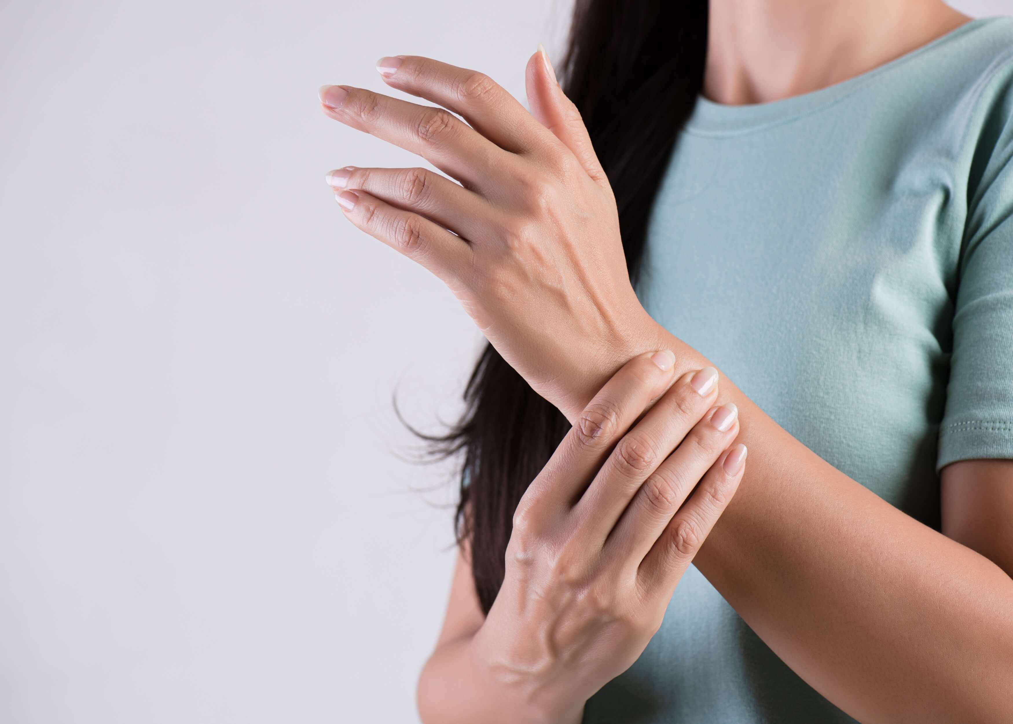
Hand and Wrist Examination (OSCE)
Introduction
 Greet the patient and introduce yourself
 Confirm patient details ✍
 Briefly explain the procedure in a patient friendly manner
 Get patient consent ✅
 Expose the patient’s elbows wrists and hands
 Wash hands ✋
 Check the patient is not in any pain

Look
Identify any signs that may be clinically relevant:
 Muscle wastage: indicates lower motor neuron lesion or disuse atrophy
 Scars: indicate prior surgery 🏨
Identify any objects/equipment that may be clinically relevant:
 Prescriptions: indicate recent medications 💊
 Aids/adaptations: e.g. wrist splints
Inspect the dorsal aspect of the hand with palm facing downwards:
 Hand posture: abnormalities can indicate pathology ✋
 Skin colour: e.g. soft tissue erythema can indicate joint sepsis or cellulitis
 Scars: indicative of prior surgery/trauma 🏨
 Swelling: compare hands and wrists
 Z-thumb: indicates rheumatoid arthritis
 Muscle wastage: due to lower motor neuron pathology or chronic joint pathology
 Heberden’s nodes: associated with osteoarthritis, identified at the distal interphalangeal joint (DIPJ) ✋
 Bouchard’s nodes: also associated with osteoarthritis, identified at the proximal interphalangeal joints (PIPJ) ✋
 Swan neck deformity: associated with rheumatoid arthritis, identified at the DIPJ (DIPJ flexion and PIPJ hyperflexion) ✋
 Boutonnières deformity: also associated with rheumatoid arthritis, PIPJ flexion and DIPJ hyperextension ✋
 Nail pitting and onycholysis: indicative of psoriasis/psoriatic arthritis
 Psoriatic plaques: scaly plaques, increases risk of psoriatic arthritis
 Splinter haemorrhages: longitudinal, red-brown haemorrhage beneath the nail caused by psoriatic nail disease/trauma/sepsis/infective endocarditis/ vasculitis
 Skin thinning/bruising: due to long term steroid use e.g. inflammatory arthritis patients
Inspect the palmar aspect of the hand with the palms facing upwards:
 Scars: indicate prior surgery or trauma 🏨
 Elbows: assess for presence of psoriatic plaques or rheumatoid arthritis
 Hand posture: identify abnormality ✋
 Dupuytren’s contracture: thickening of palmar fascia, eventually causing cords which lead to finger and thumb deformity
 Swelling: identify by comparing hands and wrists
 Thenar/hypothenar wastage: indicates median nerve damage (e.g. carpal tunnel syndrome)
What are the different types of arthritis?
- Osteoarthritis: most common, joint pain made worse by activity, causes inflammation, loss of cartilage and the adjacent bone is remodelled ✋
- Symptoms: Heberden’s nodes, Bouchard’s nodes, crepitus and reduced joint movement
- Rheumatoid arthritis: autoimmune disease, causes synovial joint inflammation, destruction of periarticular tissue etc. ✋
- Symptoms: joint pain/swelling/stiffness in the morning, symmetrical hand inflammation, muscle wastage, Z-thumb, Swan neck and Boutonnières deformities, and ulnar deviation
- Psoriatic arthritis: autoimmune disease associates with psoriasis ✋
- Symptoms: joint and tendon inflammation, joint pain and digit swelling
Feel
Assess the hands with the palms facing upwards:
 Assess temperature: use the back of your hands for comparison between the hands ✋
- Raised temperature in conjunction with selling/tenderness, can indicate inflammatory/septic arthritis
 Assess the radial and ulnar pulse: for confirmation that the hand’s blood supply is adequate
 Assess thenar and hypothenar eminence muscle bulk: wastage may be due to lower motor neuron lesions or disuse atrophy
 Assess palmar thickening: palpation of the palm can identify thickened palmar fascia bands which are associated with Dupuytren’s contracture ✋
 Assess sensation of the median and ulnar nerves:
- Median is assessed over the index finger and thenar eminence 👆
- Ulnar is assessed over the little finger and hypothenar eminence 👆
Assess the hands with palms facing downwards:
 Assess the sensation of the radial nerve: over the first dorsal webspace
 Assess the temperature: compare the temperature of the joints and the elbow 💪
 Squeeze the metacarpophalangeal joint (MCPJ): observe patient for any indication of discomfort or tenderness which indicates active inflammatory arthropathy ✊
 Bimanually palpate hand joints: identify and compare tenderness, temperature and irregularity (DIPJ, PIPJ, MCPJ and metacarpophalangeal joint [CMCJ])
 Palpate the anatomical snuffbox: assess for tenderness which indicates a scaphoid fracture (when you fall of an outstretched hand)
 Bimanually palpate the wrist: identify tenderness or joint line irregularities
Palpate the elbows 💪:
- Begin at the ulnar border, palpating to the elbow
- Identify any tenderness, psoriatic plaques or rheumatoid arthritis

Move
Assess active (independently controlled) movement:
- Open fist and extend finger
- Form a fist ✊
- Extend wrists with palms facing each other (normal = 90°) 📐
- Flex wrists completely with back of hands facing each other (normal = 90°) 📐
Assess passive (clinician controlled) movement:
 As you move the joint, ensure you feel the crepitus, observing for any discomfort/restriction ✋
 Ensure the patient is fully relaxed beforehand and is aware they should not feel any pain
 Repeat steps 1-4 when assessing active movement, but passively this time
Assess motor function:
 Radial nerve assessment: extend wrist and fingers against resistance
- Assessing: wrist and finger extensors
 Ulnar nerve assessment: abduct index finger against resistance
- Assessing: first dorsal interosseous
 Median nerve assessment: abduct thumb against resistance
- Assessing: abductor pollicis brevis
Function
Assess the patient’s hand function:
 Power grip: Ask the patient to squeeze your fingers ✊
 Pincer grip: ask the patient to squeeze your finger between your index finger and thumb
 Small object: ask the patient to pick up a small object such as a coin
Special tests
Tinel’s Test:
 Tap over the carpal tunnel to identify median nerve compression
 Used to help diagnose carpal tunnel syndrome when the patient develops a tingling in the thumb and radial
Phalen’s Test:
 Used to support diagnosis of suspected Carpal tunnel syndrome
 Ask patient to put the backs of their hands together, holding their wrists in maximum forced flexion for 1 minute ⌛
 Recurrence of carpal tunnel syndrome symptoms is a positive result
Carpal tunnel syndrome:
 Due to compression of the median nerve in the carpal tunnel, causing pain, paraesthesia and grip weakness
Completion
 Tell the patient the examination is complete ✅
 Thank patient
 Wash hands ✋
 Summarise what the examination revealed
Summary:
- Greet the patient and briefly explain the examination
- Inspect the patient to identify anything clinically relevant
- Inspect the dorsal and palmar aspects of the hand
- Assess the hands with the palms facing upwards and then downwards
- Palpate the elbows
- Assess active and passive movement of the hands and wrist
- Assess motor function
- Assess hand function
- Perform special tests such as Tinel's and Phalen's test, and assess for Carpal Tunnel syndrome
Related Articles
This step by step guide is designed to take you through the ankle and foot examination in OSCEs.
This step by step guide is designed to take you through the GALS examination in OSCEs.


-250x250.jpg)


