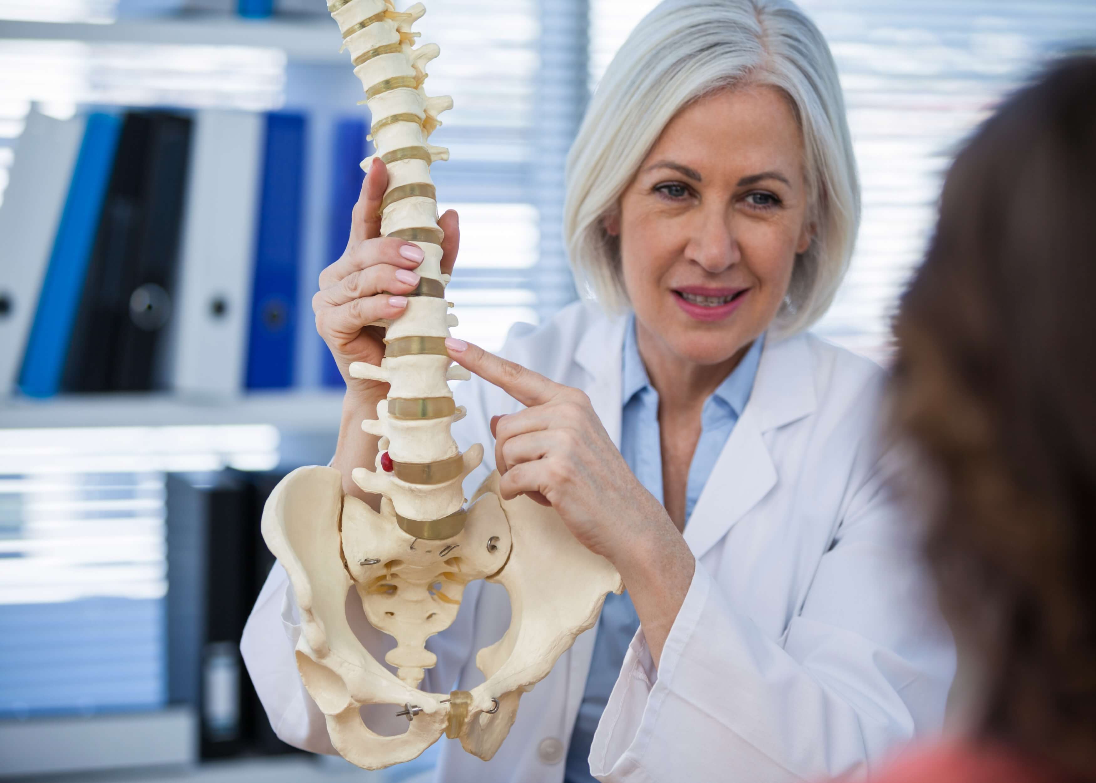
Spine examination (OSCE)
Introduction
 Greet the patient and introduce yourself
 Confirm patient details
 Get patient consent ✍
 Expose the required region and ask the patient to stand
 Wash hands ✋
 Check the patient is not in any pain
Look
Inspect the patient for any clinically relevant signs:
 Scars: indicate prior spinal surgery 🏨
 Obesity: joint pathology risk factor
 Muscle wastage: indicates disuse atrophy
Inspect for any objects/equipment that may be clinically relevant:
 Prescriptions: indicate recent medications 💊
 Aids/adaptations: wheelchairs/walking aids ♿
Inspect the anterior aspect of the spine:
 Posture: asymmetry may indicate scoliosis/joint pathology
 Lateral pelvic tilt: caused by scoliosis/hip abductor weakness/different leg lengths
 Scars: indicate prior surgery/trauma 🏨
 Shoulder girdle asymmetry: caused by scoliosis/ fractures/dislocation/arthritis
Inspect the lateral aspect of the spine:
 Loss of normal lumbar lordosis: indicates sacroiliac joint disease
 Cervical lordosis: hyperlordosis indicates chronic degenerative joint disease
 Thoracic kyphosis: (normal = 20-45°), hyperkyphosis indicates Scheuermann’s disease 📐
Inspect the posterior aspect of the spine:
 Bruising: indicates recent traums/surgery
 Spinal alignment: lateral curvature indicates scoliosis
 Muscle wastage: wastage of paraspinal muscles due to pathology/reduced mobility
 Iliac crest alignment: misalignment can be caused by different leg lengths or weakness of the hip abductor muscles
 Abnormal hair growth: due to abnormalities such as spina bifida
Observe the patient as they walk to the end of the room and back:
 Gait cycle: identify abnormalities 🚶
 Waddling gait: indicates weakness of the hip abductor muscles on both sides, associated with myopathies
 Trendelenburg’s gait: indicates weakness of the hip abductor muscles on one side, due to L5 radiculopathy or superior gluteal nerve lesion
 Leg length: differences can indicate joint pathology
 Limp: can indicate joint pain/weakness
 Slow turning: can indicate joint restrictions
 Footwear: unequal wearing of the sole can indicate an abnormal gait
 Movement: reduced range of movement indicates chronic joint pathology
Normal gait cycle:
- Heel makes contact with floor 🚶
- Foot becomes flat and weight is transferred onto it
- Weight balanced on flat foot’s leg
- Heel lifted off floor
- Toes lifted off floor
- Foot swings forward and cycle begins again
Feel
 Palpate the spinal processes and sacroiliac joints to assess alignment and identify any tenderness
 Palpate the paraspinal muscle to identify any tenderness or muscle spasms

Move
 Assess the following movement actively (independently controlled) and where abnormalities are observed, repeat passively (clinician controlled)
Cervical spine movements:
 Flexion: ask patient to touch chin and chest (normal = 0-80°) 📐
 Extension: ask patient to look to ceiling (normal = 0-50°) 📐
 Lateral flexion: ask patient to touch ear and shoulder on same side (normal = 0-45°) 📐
 Rotation: ask patient to turn head left and right (normal = 0-80°) 📐
Lumbar spine movements:
 Flexion: ask patient to keep legs straight and touch toes
 Extension: ask patient to lean back as far as possible (normal = 10-20°) 📐
 Lateral flexion: ask patient to slide down outer aspect of both sides, one after the other, keeping legs straight
Thoracic spine movements:
 Rotation: ask patient to sit on side of bed with arms crossed across chest, then turn to left and right as far as possible
Special tests
Schober’s test:
 Used for identification of restricted flexion of lumbar spine e.g. in ankylosing spondylitis
- Mark skin 5cm below and 10cm above the posterior superior iliac spine on both sides
- Ask patient to touch their toes
- Measure distance between the 2 lines when they bend
 Normal: distance should increase from 15cm to 20cm 📏
 Reduced distance: indicates pathology e.g. ankylosing spondylitis
Sciatic stretch test:
 Used for identification of sciatic nerve irritation
- Hold patient’s ankle as they are lying down
- Raise the leg from the hip (normal = 80-90° movement range) 📐
- When hip is flexed, point the foot towards the shin
 Pain in the posterior thigh/buttocks: positive result, indicating sciatic nerve irritation
Femoral nerve stretch test:
 Used for identification of femoral nerve irritation
- Flex patient’s knee to 90° 📐
- Extend hip joint
- Extend the ankle, pointing toes/feet
 Pain in thigh and/or inguinal region: positive result
Completion Summary:
 Tell the patient the examination is complete ✅
 Thank patient
 Wash hands ✋
 Summarise what the examination revealed
Summary:
- Greet the patient and briefly explain the examination
- Inspect the patient to identify anything clinically relevant
- Inspect the anterior, lateral and posterior aspects of the spine
- Observe the patient walking to the end of the room and back to assess whether their gait is normal
- Palpate the spine to identify any tenderness
- Assess cervical, lumbar and thoracic spine movements
- Perform special tests such as Schober's test, Sciatic stretch test and femoral nerve stretch test
- Complete the examination by thanking the patient
Related Articles
This step by step guide is designed to take you through the GALS examination in OSCEs.


-250x250.jpg)


