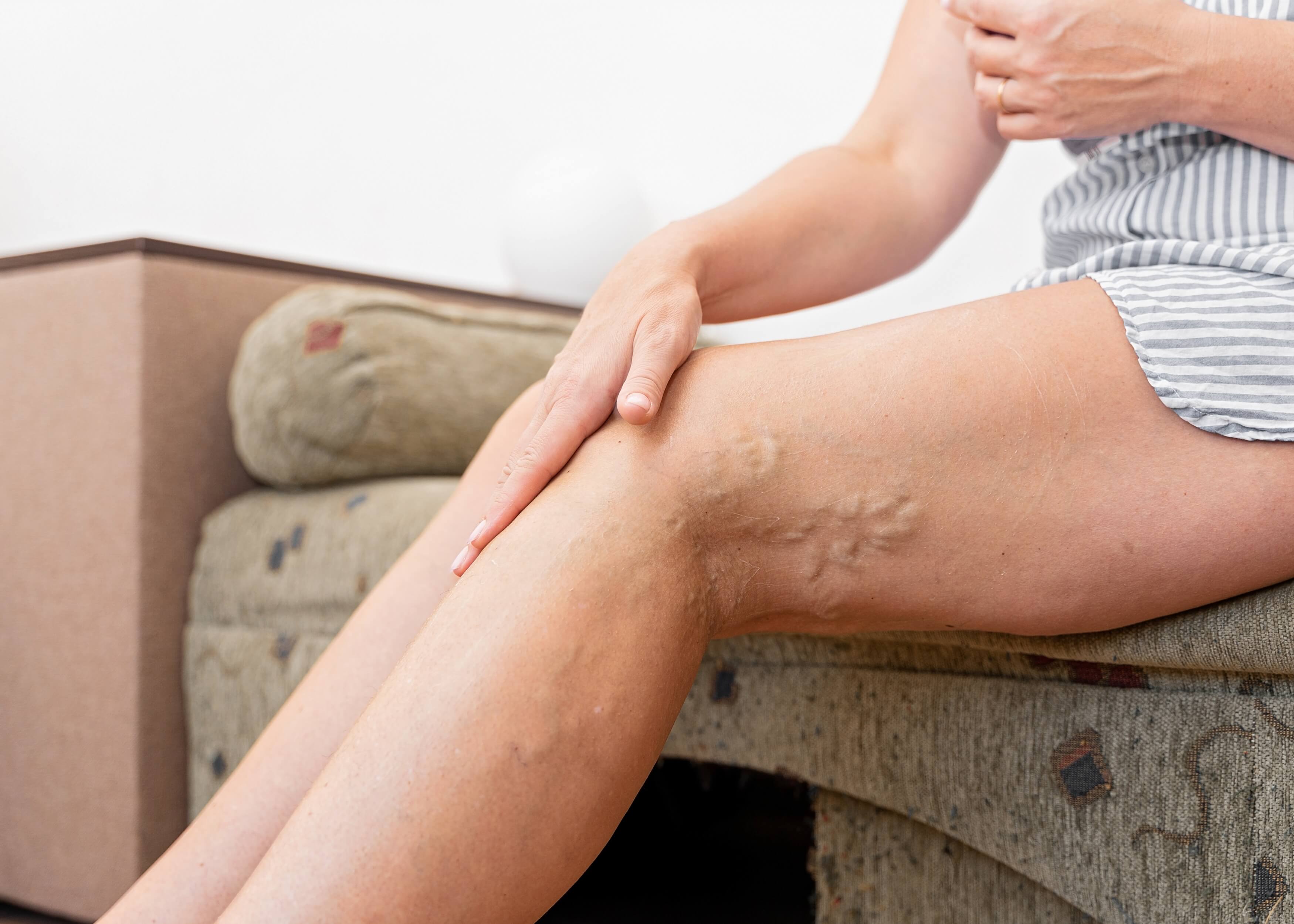
Varicose Vein Examination (OSCE)
Introduction
- Greet the patient and introduce yourself
- Confirm patient details ✍
- Explain the procedure in a patient friendly manner 👨
- Explain that a member of staff from the ward will be present throughout the procedure as a chaperone
- Get patient consent ✅
- Wash hands ✋
- Ensure the patient is standing with their lower limbs exposed
- Check the patient is not in any pain
General Inspection of the patient
Identify any clinically relevant signs:
➖ Ulcers: can indicate diseased veins/arteries
➖ Scars: can indicate that the patient has had previous ulcers which are now healed, or previous surgery
Identify and objects or equipment that may provide an insight into their medical status:
➖ Mobility aids: indicates their current mobility ♿
➖ Vital signs: indicates their current clinical status
➖ Prescriptions: indicates any relevant medications they are on 💊
➖ Medical Equipment: for example, dressings on wounds or compression stockings
Inspection of patient’s legs
Surgical scars: clarify the surgery with patient, for example, a low groin scar can indicate venous treatment (remember venous treatment is minimally invasive nowadays and so you won’t see any scars)
Venous eczema: inflammation caused by fluid in the tissues due to venous hypertension (you can ask “is it itchy” to help identify it!)
➖ Lipodermatosclerosis
➖ Atrophie-blanche (white atrophy): depressed atrophic plaques which are star shaped and ivory-white with red dots and surrounding hyperpigmentation
➖ Crusty plaques which are blistered and itchy or dry and scaly plaques
➖ Orange-brown pigmentation patches
Lipodermatosclerosis: a form of panniculitis caused when the innate immune system is activated in soft tissues for a prolonged period. It often occurs in individuals with chronic venous insufficiency (CVI) which is often caused by varicose veins.
➖ Clinical features of Lipodermatosclerosis:
➖ Swelling
➖ Hyperpigmentation
➖ Skin hardening or thickening
➖ Erythema
➖ ‘Tapered’ appearance of legs above the ankles
Venous ulcers: believed to be caused when the venous valves do not function properly
➖ Mild pain
➖ Shallow depth
➖ Large, irregularly shaped border
➖ Commonly located at the ankle’s medial aspect
Saphena varix: when the saphenous vein is dilated in the groin where it meets the femoral vein
➖ Lump (2-4cm inferior lateral to pubic tubercle)
➖ Bluish colouration of lump
➖ Lump is soft to palpate
➖ Lump disappears when patient is lying down
Arterial disease: it is important to assess for this when treating issues of the venous system as it may mean the patient is unsuitable for the standard varicose vein treatment of compression therapy as it may put them at risk of secondary ishcaemia. Similarly, if the patient suffers from venous ulcers, it is important to ensure their arterial supply is adequate before you treat it
➖ Grangrene
➖ Hair loss
➖ Arterial ulcers
➖ Peripheral cyanosis
➖ Lower temperature ❄
➖ Peripheral pallor
Varicose veins: dilated superficial veins, commonly seen on the legs, however when found on the genitals or buttocks, it can indicate pathology of the venous system within the pelvis
Great saphenous vein: this is the largest vein in the human body, it runs the entirety of the way up the medial side of the leg
- It originates where the big toe’s dorsal vein meets the dorsal venous arch of the foot ?
- It then passes in front of the medial malleolus before running up the leg on the medial side
- When it reaches the knee, it passes over the posterior border of the femur bone’s medial epicondyle
- Finally, after passing through the proximal anterior thigh, it joins the femoral vein
Small saphenous vein: it drains the lateral side of the lower leg
- It originates where the dorsal vein of the fifth digit meets the dorsal venous arch of the foot 👣
- It runs along the lateral aspect of the foot followed by the posterior aspect of the legs
- It drains into the popliteal vein at or above knee joint level (the saphenopopliteal junction)

Assess varicosities
Assess the temperature of varicosities: using the back of your hand
➖ Higher temperature 🌡 indicates inflammation
Palpate varicosities: palpate the entire length, asking the patient when they feel pain
➖ Tenderness and redness indicate phlebitis
➖ Tenderness and hardness indicate thrombophlebitis
Additional lower limb assessment
➖ Assess the limb for pitting oedema: moving up the leg, use your fingertis to apply pressure for a few seconds above the medial malleolus, then observe whether an indentation is formed
➖ Cause: heart failure 💔 (its presence can also impact the integrity of the skin)
➖ Palpate lower limb pulses to assess the arterial blood supply of each leg: moving in a proximal to distal direction (use a Doppler if not palpable)
➖ Femoral pulse 💓: palpated at the mid-inguinal point to check pulse is present and check the volume
➖ Popliteal pulse 💓: palpate in the inferior region of the popliteal fossa
- When the patient’s legs are relaxed, place your thumbs on the tibial tuberosity
- Flex the knew to 30° as you place your fingers in the popliteal fossa to feel the pulse
➖ Posterior tibial pulse 💓: palpated posterior to the tibia’s medial malleolus, compare strength of pulse between the two feet
➖ Dorsalis pedis pulse 💓: palpated over the dorsum of the foot, lateral to the extensor hallucis longus tendon, or over the 2nd and 3rd cuneiform bones. Comparison of the strength of this pulse between the two feet should be made
Percussion ‘tap test’:
➖ A rarely used method to assess the venous valve competency of lower limbs
- Apply a small amount of pressure onto the saphenofemoral junction with one finger 👉
- Tap the varicose vein (lower down the leg) being assessed
- If you detect a ‘thrill’ with your finger, it indicates that the venous valves are incompetent causing vein continuity, as the valves did not prevent the thrill
Auscultation:
➖ Another rarely used procedure which involves placing your stethoscope’s bell over the varicosity, listening for a bruit which indicates turbulent blood flow which can suggest arteriovenous malformation
Additional tests
Venous duplex scanning:
➖ Today, all patients being considered for varicose vein treatment should undergo venous duplex scan of the whole superficial venous system for the following reasons:
➖ To confirm where the incompetence originated (e.g. saphenofemoral or saphenopopliteal junction)
➖ To assess whether the veins are straight enough to undergo endovenous treatment
➖ To establish the role the deep venous system is playing (e.g. the patient may be at risk of chronic limb swelling if the superficial venous system is treated when it is returning venous blood because the deep veins are incompetent)
Handheld Doppler:
➖ A traditional test rarely performed since the advent of venous duplex scanning
Trendelenburg test/ Tourniquet test:
➖ To locate the incompetent venous valves
- Lift leg upwards as patient lays flat and milk the leg towards the groin area to empty the superficial veins
- Place tourniquet over the saphenofemoral junction
- Observe the veins filling as the patient stands
- No filling and veins remain collapsed: incompetent venous valves
- Veins fill: incompetent valve is inferior to the saphenofemoral junction
- Repeat steps 1-4 but this time place tourniquet 3cm lower than the first time
- Repeat until you have identified the location of the incompetent valves, visualised by observing where filling stops
Cough impulse test:
- Ask patient to cough with your hand over the saphenofemoral junction
- If you feel an impulse, this is indicative of a saphena varix
Perthe’s test:
Allows you to distinguish between venous valvular insufficiency in the deep perforator and superficial venous system
- With the patient standing, apply tourniquet at mid-thigh level
- Ask patient to walk around for 5 minutes
- If the varicose vein is less distended after 5 minutes, it indicates that the deep venous valvular system is sufficient, suggesting the problem is with the superficial veins
- If the varicose veins remain distended after 5 minutes, it indicates there is a problem with the deep venous system, potentially caused by deep vein thrombosis
Completion of the Examination
- Explain to the patient that the examination is complete, and thank them
- Wash hands ✋
- Summarise what the examination revealed
Summary:
- Greet the patient, introduce the chaperone and briefly explain the procedure
- Inspect the patient for any clinically relevant signs
- Inspect the patient's legs for any clinically relevant signs
- Assess varicosities
- Assess lower limbs e.g. measure pulses, perform percussion tap test and auscultation
- Perform additional tests: Venous duplex scanning, Handheld Doppler, Trendelenburg test, Cough impulse test and Perthe's test
- Complete the examination by thanking the patient
Related Articles
This step by step guide is designed to take you through the peripheral vascular examination in OSCEs.


