Your shopping cart is empty!


PERSONAL BLOG
What is Addison's Disease?
All You Need to Know about Addison’s Disease 👩🏻🎓
Addison’s disease is a condition that can become an emergency if not promptly treated. This article describes the incidence, aetiology, risk factors, pathophysiology, clinical presentation, investigation, management, differential diagnosis and provides patient advice.
What is Addison’s Disease? ❓
Addison’s disease, which is also referred to as primary adrenal insufficiency, is a disease that occurs as a result of the destruction of the bilateral adrenal cortex, thereby leading to a decrease in the production of adrenal cortex hormones.
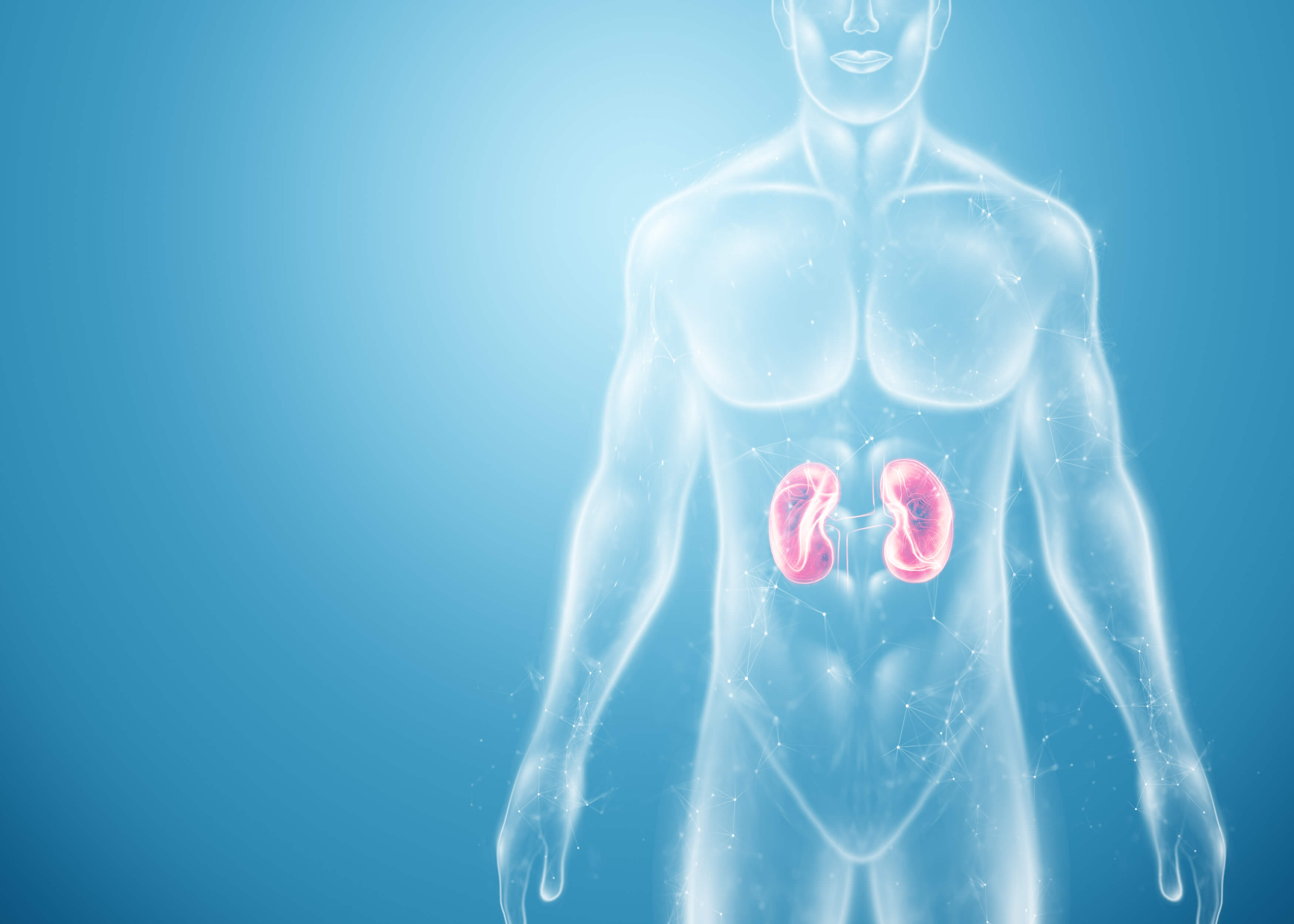
The adrenal glands, which are located just above both kidneys are small triangular-shaped glands, they produce hormones in response to the brain, or more specifically, in response to the hypothalamic-pituitary axis (HPA).
The adrenal gland comprises an outer cortex and an inner medulla. Adrenocorticotropic hormone (ACTH), which is a hormone secreted by the anterior pituitary gland, stimulates the adrenal cortex to produce 3 hormones, namely:
- Mineral corticoids (aldosterone)
- Glucocorticoids (cortisol)
- Androgens
Incidence/Epidemiology 👨👩👧👦
Addison’s disease is a relatively rare disease that has an incidence of about 0.6 per 100,000 of the population. It is a condition that ranges with a prevalence of 4 to 11 in 100,000 of the general population per person.
- The common age of presentation is around 30 to 50 years of age, although it can occur at any age.
- It is more common in women than men.
Most often, the insidious nature of the disease leads to a delay in the diagnosis of the patient until an acute adrenal crisis develops, which is a potentially life-threatening emergency.
Aetiology ⚠️
Adrenal insufficiency can be classified as primary or secondary.
Primary Adrenal Insufficiency is caused by the inability of the adrenal cortices to produce sufficient adrenocortical hormones, so in other words, any disease process that brings about direct injury to the adrenal cortex can result in Addison’s disease.
The main and most common cause of primary adrenal insufficiency is autoimmune (70%). Here, auto-antibodies are being produced against adrenal cortex cells as well as 2-hydroxylase enzyme within the adrenal gland.
This autoimmune destruction can either be an isolated finding or an autoimmune polyglandular endocrinopathies (type 1 and 2). Isolated autoimmune adrenalitis accounts for 30-40%, whereas 60-70% develop adrenal insufficiency as part of autoimmune polyglandular syndromes (APS).
- In countries with a high prevalence of HIV/AIDS, tuberculosis is an increasing cause.
- Other infections include sepsis, cytomegalovirus, fungal infections, syphilis etc.
- Infiltration diseases e.g. haemochromatosis, amyloidosis.
- Adrenal cortex malignancy (secondary metastases from the lung for example).
- Haemorrhage infarction.
- Latrogenic causes (the surgical removal of the adrenal gland).
- Medications: certain drugs can lead to decreased cortical production and cause adrenal insufficiency. E.g. etomidate, mitotane, aminoglutethimide.
- Rare – congenital adrenal hyperplasia which presents with ambiguous genitalia and adrenal failure.
Secondary Adrenal Insufficiency normally arises from:
- A hypothalamic-pituitary disease where there is inadequate production of ACTH.
- Use of long term steroid therapy that leads to hypothalamic-pituitary-adrenal suppression.
In Conclusion
- Primary – caused by an autoimmune-mediated adrenal gland dysfunction. Here, both cortisol and aldosterone are reduced.
- Secondary – chronic glucocorticoid administration or use that result in hypothalamic-pituitary dysfunction. Here, there is only cortisol deficiency.
The following factors and disease processes increase the likelihood of getting Addison’s disease:
- Female gender
- Tuberculosis infection
- Meningococcal infection
- Adrenal haemorrhage
- Auto-immune diseases like Type I diabetes, pernicious anaemia, Graves’ disease, Hypoparathyroidism, Vitiligo, Hypopituitarism.
Pathophysiology 🔎
Normally, the corticotropic releasing hormone stimulates the cells within the anterior pituitary gland to secrete adrenocorticotropic hormone (ACTH), and this ACTH will enter the circulation and travel towards the adrenal gland or more specifically the adrenal cortex.
An increase in cortisol in the circulation will have negative feedback on the hypothalamus, it will tell the hypothalamus to stop producing corticotropic releasing hormone (CRH), thereby decreasing the stimulation of ACTH.
However, in Addison’s disease, there is a loss of this negative feedback on the hypothalamus and the opposite happens, so because of the destruction of the adrenal cortex, there is reduced production of cortisol, followed by that of aldosterone, which eventually results in the elevation of ACTH and CRH production.
There is also an elevation in melanocyte-stimulating hormone (MSH) and it is believed that ACTH binds to the melanocyte receptors which are responsible for skin pigmentation.
Clinical Presentation
A patient with Addison’s disease would present with the following symptoms:
- Tiredness/fatigue
- Dizziness
- Weight loss
- Nausea and vomiting
- Diarrhoea
- Abdominal pain
- Decreased libido in women
- Anorexia
- Depression
- Arthralgia (joint pain) and Myalgia (muscle pain)
- Syncope from postural hypotension
- Generalized hyperpigmentation is more pronounced in skin areas that are exposed to increased friction or sunlight e.g. the elbows, knuckles and palmar creases.
- On Examination, a patient with Addison’s disease will present with low-grade fever, signs of dehydration, loss of hair, cachexia (a wasting disorder that causes extreme weight loss and muscle waste and can include the loss of body fat), and postural hypotension.

Hyponatremia (low serum sodium) is a very characteristic biochemical feature in primary adrenal insufficiency and is found in about 80% of patients at presentation.
Hyperkalaemia (a high level of potassium) is also present in 40% of patients at the initial diagnosis.
In Summary,
- Signs and symptoms such as fatigue and loss of energy are relatively nonspecific, and most often results in delay or misdiagnoses (e.g. as depression or anorexia).
- A distinctive feature of primary adrenal insufficiency is hyperpigmentation, which happens because of the stimulation of melanocytes as a result of excess ACTH.
Addisonian Crisis 🚑
Addisonian crisis, also called acute adrenal crisis, is a life-threatening emergency that usually occurs after a prolonged period of non-specific symptoms due to loss of both glucocorticoids and mineral corticoid secretion.
The diagnosis is only made after the patient presents with the following features:
- Hypotension
- Hyponatremia
- Hyperkalaemia
- Hypoglycaemia
- Acute adrenal crisis is often triggered or brought about by an intercurrent illness, stress, accident or surgery.
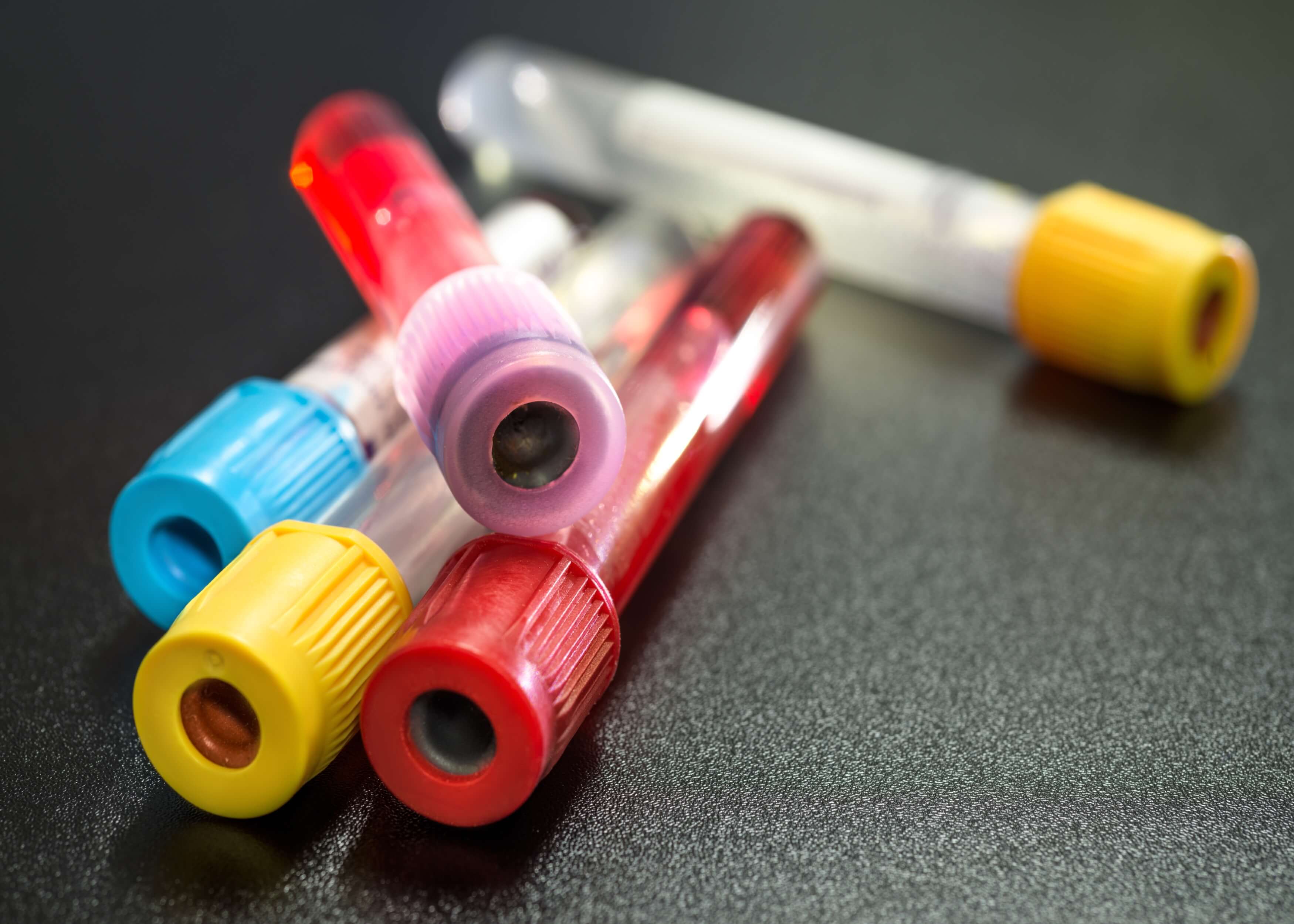
Investigations Used to Diagnose Addison’s Disease 👨⚕️
ACTH Level and Corticotropin Stimulation Test
- In uncertain cases, ACTH (cosyntropin) stimulation test can be used as a way of confirming the diagnosis.
- The short cosyntropin stimulation test is a safe and reliable test with exceptional predictive value.
- Plasma cortisol levels are normally measured at 0, 30 and 60 minutes after the administration of ACTH.
- A high ACTH level is present which causes the further rise after the stimulation of CRH but leads to a reduced level of serum cortisol.
It is important to remember that:
- With primary adrenal insufficiency, there is elevated ACTH.
- In secondary adrenal insufficiency, ACTH is low and does not respond to CRH.
With ACTH Stimulation
- If it is normal, it would show an adequate response and a peak cortisol level of >18 mcg/dL.
- However, with adrenal insufficiency, there is less response or no response at all (an absent or impaired cortisol response).
Single Cortisol Measurements
- A random cortisol level is carried out and this would typically show a low cortisol level.
- Normally, cortisol follows a diurnal (during the day) pattern where it is highest in the morning so therefore, an early morning level should be taken.
- Single cortisol measurements are of little value as they are unable to assess adrenal function, although a low random cortisol level of <3 mcg/dl during the day is highly suggestive of primary adrenal insufficiency.
Aldosterone and Renin Level
Serum aldosterone and renin levels must be taken to know whether mineral corticoid deficiency is present.
- Plasma renin activity is high as a result of the low serum aldosterone in primary adrenal insufficiency.
- In secondary adrenal insufficiency, the serum aldosterone level will be normal.
Anti-21-Hydroxylase Antibodies
- Anti-adrenal antibodies which are also known as 21-hydroxylase antibodies are important markers of autoimmune destruction of the adrenal cortex.
- During the initial presentation of primary adrenal insufficiency, it's important these patients undergo screening for steroid autoantibodies as it is a marker for autoimmune adrenalitis.
- These antibodies help to determine the cause of Addison’s disease.
TSH Level
- In adrenal insufficiency, there may be a slight elevation of thyroid-stimulating hormone (TSH) level.
- This can occur due to the reduction of cortisol levels and an abnormal TSH circadian rhythm.
- However, it is also important to always consider hypothyroidism as a likely diagnosis in cases where there is a persistent TSH elevation.
Laboratory Findings
The lab findings typically seen in primary adrenal insufficiency are hyponatremia, hyperkalaemia, hypoglycaemia.
- Hyponatremia (low sodium level in the blood) is caused by both cortisol and aldosterone deficiency.
- When there is low aldosterone in the blood, there is increased sodium waste and reduced water retention due to the diminished inhibition of antidiuretic hormone (ADH) release by cortisol.
- Hyperkalaemia (high potassium level in the blood) due to low aldosterone levels cause the retention of potassium in the blood.
- A way to distinguish primary from secondary is that hyperkalaemia occurs only in primary adrenal insufficiency.
- Blood glucose levels may be low due to hypoglycaemia. Hypoglycemia is more frequently seen in children.
- A full blood count may also reveal neutropenia (decreased circulating neutrophils), lymphocytosis (high lymphocyte count in the blood) and eosinophilia (a higher than normal level of eosinophils).
Imaging Techniques
- Chest and abdominal X-rays may show evidence of tuberculosis or calcified adrenal glands as a cause of Addison’s disease.
- An abdominal CT scan is important and may reveal adrenal haemorrhage, adrenal infiltration and adrenal tumours or masses.
Ways to Treat and Manage Addison’s disease
- It is very important for primary adrenal insufficiency to be recognized early and treated immediately as the failure to do this results in an adrenal crisis which is a severe endocrine emergency.

Maintenance Therapy 💊
- The patient is required to take a life-long treatment with hormonal replacement.
- This maintenance therapy aims to provide a replacement to maintain a physiologic level of glucocorticoid and mineralocorticoid.
- Glucocorticoids: hydrocortisone 5 to 25 mg/day (in 2 or 3 divided doses) so hydrocortisone is given at 20-30 mg daily (e.g. 10 mg on waking, 5 mg at 12:00 hours, 5 mg at 18:00 hours).
- OR prednisone 3 to 5 mg daily
- Mineralocorticoids: fludrocortisone 0.05 to 0.2 mg daily.
- In children, hydrocortisone is given initially at 8 mg/m²/day orally, divided into 3 or 4 doses.
Patients With an Adrenal Crisis Would Require the Following treatment: 💉
- Resuscitate the patient with fluids to restore their intravascular volume and this is done with intravenous normal saline.
- 1 litre of 0.9% saline should be given over 30-60 mins with 100 mg of intravenous bolus of hydrocortisone.
- Correct the hormone deficiency which is both glucocorticoids and mineral corticoids.
- Hydrocortisone is administered for the immediate treatment of hormone deficiency.
- The initial dose is 100 mg IV bolus, followed by 50 to 100 mg IV every 6 hours over 24 hours.
- In children, 50 mg/m² (max 100 mg) IV bolus, followed by 50 to 100 mg/m².
- Mineralocorticoids like fludrocortisone are not necessary during the acute phase because the high cortisone doses provide sufficient mineralocorticoid activity. It should be introduced later on.
- Dextrose is given to correct hypoglycaemia. It should be treated promptly so glucose (5% glucose in isotonic saline) should be infused if there is hypoglycaemia.
- Oral replacement medication is then started, unless the patient is unable to take oral medication, therefore, initially, hydrocortisone 20 mg is given, every 8 hours over 24 hours, then it is reduced to 20-30 mg in divided doses over the next few days.
- These doses should always be adjusted according to the patient’s clinical response and when electrolyte abnormalities become normal.
- The lowest possible dose should always be given first to control symptoms and to minimize the side effects of the drugs.
- Plasma renin levels are used to adjust the doses. Fludrocortisone is delivered to the patient at a sufficient dose so that the plasma renin level is kept in the reference range.
Differential Diagnosis
The differential diagnosis of Addison’s disease includes diseases or conditions that mimic a few signs and symptoms of adrenal insufficiency.
- Shock
- Sepsis
- Hypothyroidism
- Chronic fatigue syndrome
- Anorexia
Patient Advice and Education 📚
It is important for a patient with Addison’s disease to understand their illness and should receive education about the management and monitoring of primary adrenal insufficiency.
All patients that require replacement steroids should know:
- How to increase their steroid replacement dose for any intercurrent illness.
- Always carry and have a "steroid" card on them.
- Wear a Medic-Alert bracelet because it gives information about their condition so that emergency replacement therapy can be given if they are found unconscious.
- They should be advised to include adequate sodium in their diet, improve their weight and monitor their blood pressure.

Summary
- Addison’s disease is a life-threatening disease that occurs as a result of adrenal gland destruction.
- Most common cause is autoimmune adrenocortical insufficiency.
- The clinical presentation of Addison’s disease is nonspecific, however, hyperpigmentation is an important feature.
- The diagnosis is established by demonstrating low cortisol and aldosterone levels, high plasma renin level, and a low cortisol response with ACTH stimulation.
- In Addisonian crisis, the diagnosis and treatment should not be delayed when the suspicion of the condition is high as this can be fatal.
- Hydrocortisone is used to manage an adrenal crisis.
- The doses of glucocorticoids should be increased if fever, infection or stress is present.
- The treatment of Addison’s disease is a life-long replacement of glucocorticoids and mineralocorticoids.
By Jessica Otoide
I-Medics Ambassador
REFERENCES
Harrison Principles of Internal Medicine 19th Edition
Kumar and Clark’s clinical medicine
https://www.ncbi.nlm.nih.gov/books/NBK441994/
https://emedicine.medscape.com/article/116467-overview

Related Articles
Everything you need to know about Addison's Disease in 60 seconds!